 At Midcoast Cardiovascular Associates, Dr. Ginkel treats nearly all conditions involving the heart, veins, and blood vessels of the cardiovascular system. Using ultrasound imaging, he investigates the functionality of the venous, arterial, and vascular systems. Ultrasound imaging is both non-invasive and safe, and is one of the methods most commonly used to perform a thorough examination of those systems. Arteries and veins of the extremities, torso and neck are most often evaluated, although many vessel types and locations can be safely assessed using this method.
The venous system is a complex network of superficial veins, deep veins, and perforator (connector) veins which all need to be evaluated to formulate an appropriate treatment plan. Most people are unaware that they have a venous condition, which can cause swelling, pain, heaviness, and the appearance of spider or varicose veins. The appearance of superficial veins can be the first sign of venous disease, but while those symptoms can be an indicator of deep venous disease, that is not always the case. A bilateral venous ultrasound allows Dr. Ginkel to further examine your superficial and deep veins to explore the area and offer an accurate diagnosis before he recommends vein treatments such as endovenous RF ablation, VenaSeal, ClariVein, or microphlebectomy.
The arterial system carries nutrient-rich blood away from the heart to supply nutrients to the organs and tissues throughout the body. These vital arteries can be damaged or closed by atherosclerosis, causing serious issues for the organs and tissues they supply. Peripheral arterial disease (PAD) can appear with a variety of signs or symptoms, depending on the location and the particular organ or tissues a vessel serves. Affected arteries that send blood to the extremities may cause consistently cold hands or feet, non-healing wounds or ulcers, and in severe cases, gangrene. Diseased carotid arteries, which are responsible for sending blood to the brain, can also cause a transient ischemic attack (TIA) or stroke.
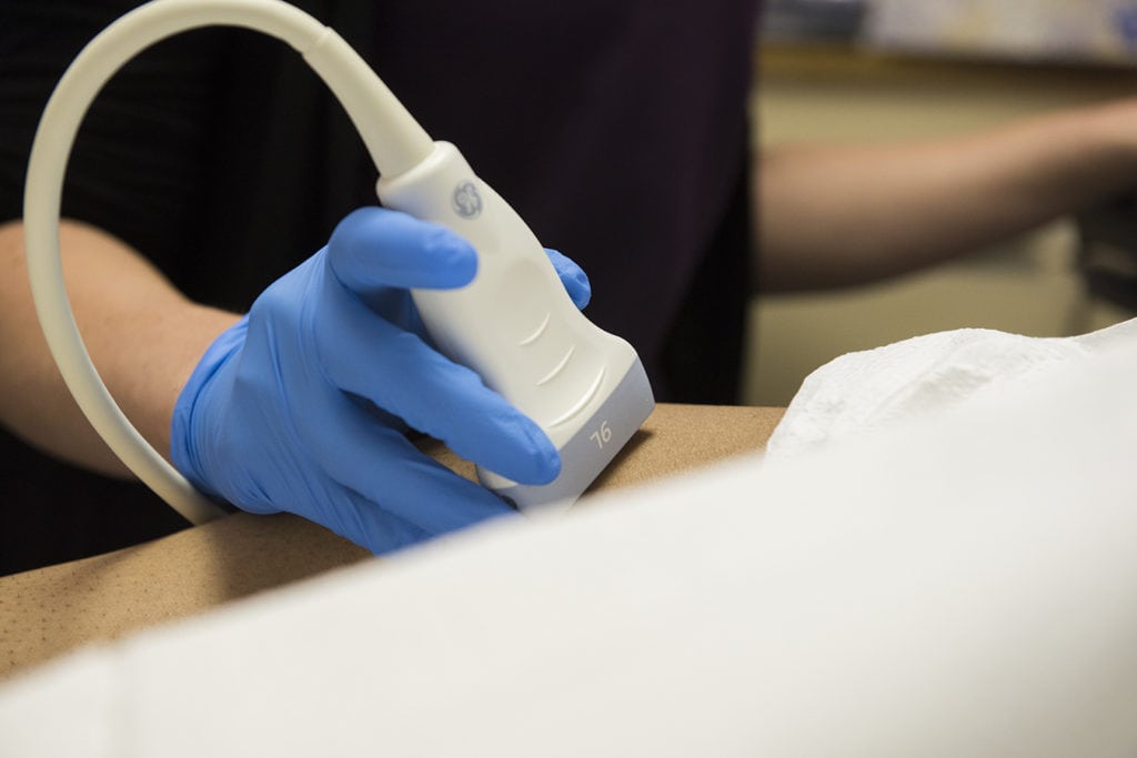
The Ultrasound Experience and What to Expect
During the ultrasound exam, a sonographer will coat the targeted area with a water-based ultrasound gel that allows the high-frequency energy to flow through the soft tissue. You’ll be positioned in various ways, depending on the vessel of interest, as the tech applies the ultrasound transducer to the area of concern. During a venous ultrasound, the tech will also work through a series of vein compression maneuvers to check for clots and insufficiencies.
Compressing the veins should be a simple process for healthy veins, however, if we’re unable to compress each vein, this could mean a blockage is present. The color (or Doppler) exam allows Dr. Ginkel to see the speed and direction at which your blood moves through the vein. If the color rushing through your veins stops in a particular area, he will be able to see these previously undetectable blockages. If the blood flows backwards, the valves within the veins are incompetent, which is known as reflux. Over time, reflux can cause edema, discoloration, and the appearance of spider veins and varicose veins.
Venous ultrasound exams have saved lives, and it’s never too early to undergo this simple procedure. Call Midcoast Cardiovascular today at 805-354-0112 to schedule a consultation with Dr. Ginkel.
Recent Comments